Figure 1C.

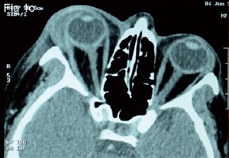
(C) CT scan of orbit shows proptosis, hypoechoic lesion with hyperechoic borders localized in preseptal tissues and anterior orbit with surrounding preseptal soft tissue swelling extending nasally and temporally

(C) CT scan of orbit shows proptosis, hypoechoic lesion with hyperechoic borders localized in preseptal tissues and anterior orbit with surrounding preseptal soft tissue swelling extending nasally and temporally