
Figure 1A.
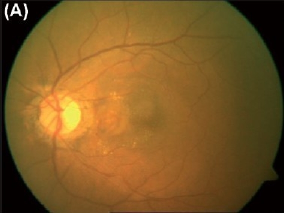
(A) Color fundus photograph of the left eye reveals a choroidal neovascular membrane underlying the papillomacular bundle. Hard exudates and subretinal hemorrhages are noted surrounding it.
Official websites use .gov
A
.gov website belongs to an official
government organization in the United States.
Secure .gov websites use HTTPS
A lock (
) or https:// means you've safely
connected to the .gov website. Share sensitive
information only on official, secure websites.
(A) Color fundus photograph of the left eye reveals a choroidal neovascular membrane underlying the papillomacular bundle. Hard exudates and subretinal hemorrhages are noted surrounding it.