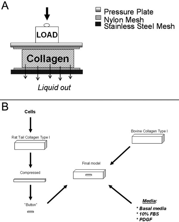
Figure 1.

A. Illustration of the collagen compression procedure is shown. Cell-seeded collagen was placed between two nylon meshes and compressed using a load for 5 minutes, during which time liquid was allowed to exit through a stainless steel mesh at the bottom. B. Schematic of the process for constructing the migration model. Cells were seeded in rat tail collagen and compressed (as shown in A), and a 6 mm button was punched out and placed inside an acellular uncompressed matrix.
