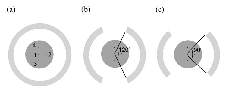
Figure 1.
Scanner setup for the (a) full, (b) 2/3 (120 degrees in-plane coverage), and (c) ½ ring (90 degrees in-plane coverage) scanners. The ring diameter and the axial length for the scanners are 15-cm. The simulated cylindrical phantom (length is 8-cm, diameter is 6 or 10-cm) has three, 5-mm diameter hot spheres (lesion 1, 2, and 3) with 8:1 uptake with respect to background, and one, 5-mm diameter cold sphere. The scanner ring diameter was fixed at 15-cm.

