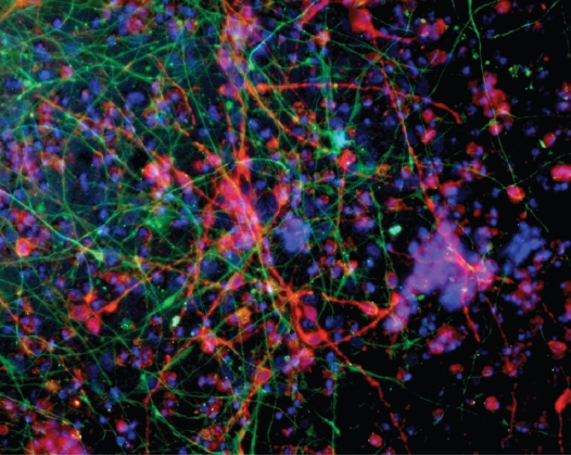
Figure 2.
Human Neural Stem Cells cultured in a RADA16-I-BMHP1 3D scaffold (3 weeks in vitro). Cell nuclei are stained with DAPI (blue), neurons with βTubulin antibody (red), and astrocytes with GFAP antibody (green). In this long-term cultures neuronal morphologies resemble fairly mature neurons. A highly connected neuronal network is shown. Branched astrocytes also give evidence of differentiation of part of the stem cell progeny toward the astroglial phenotype.

