
Fig. 1.
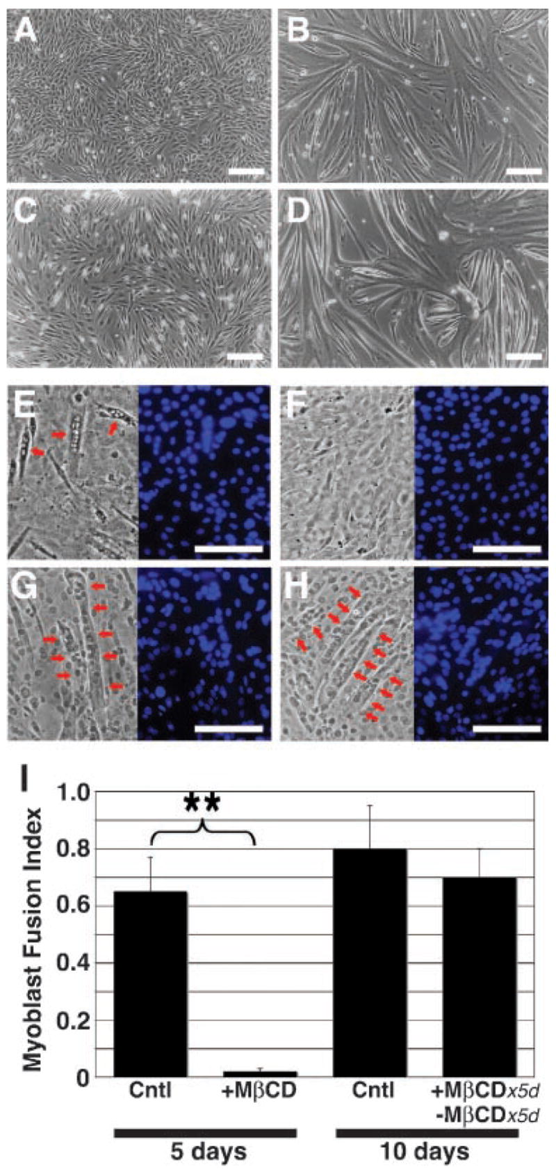
Lipid microdomains are necessary for normal myoblast differentiation. Proliferating myoblasts (A) grown under differentiation conditions formed multinucleated myotubes (B) within 5 days. C: Confluent myoblasts remained mononuclear after 5 days under differentiating conditions in the presence of MβCD. D: With subsequent washout of MβCD, the myoblasts differentiated normally within 5 days. E–G: Representative phase micrographs (left parts) of differentiating myoblasts grown under conditions as in (B–D), respectively, and stained with DAPI (blue; right parts) for calculation of MFI. H: Control myoblasts grown under differentiating conditions for 10 days. Red arrows indicate representative myotubes. A–H: Bar, 50 μm. I: Cells treated with MβCD for 5 days (see C, F) demonstrated a significantly attenuated MFI (see Materials and Methods Section) compared to untreated cells (see B,E). MFI was not significantly different between cultures grown without MβCD for 5 days (see B,E) or 10 days (see H), or for 5 days in the presence of MβCD followed by 5 days after MβCD washout (see D,G). Data shown are mean ±SEM (N =3). **P > 0.01.