
Fig. 3.
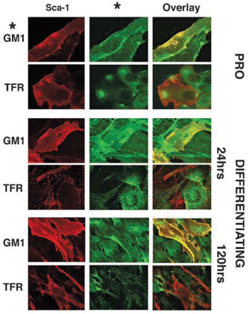
Sca-1 remains localized to lipid microdomains during differentiation. C2C12 myoblasts were imaged before (PRO) and after 24 and 120 h of differentiation. Representative data are shown. Sca-1 (red) showed overlap (yellow) with GM1 ganglioside (GM1; green), a lipid microdomain marker, in proliferating and differentiating cells. Sca-1 demonstrated minimal overlap with the transferrin receptor (TFR), a non-lipid microdomain-associated cell surface protein at all stages. These findings support the biochemical data in Figure 2, and suggest that Sca-1 constitutively localizes to lipid microdomains in myoblasts.