
Fig. 1.
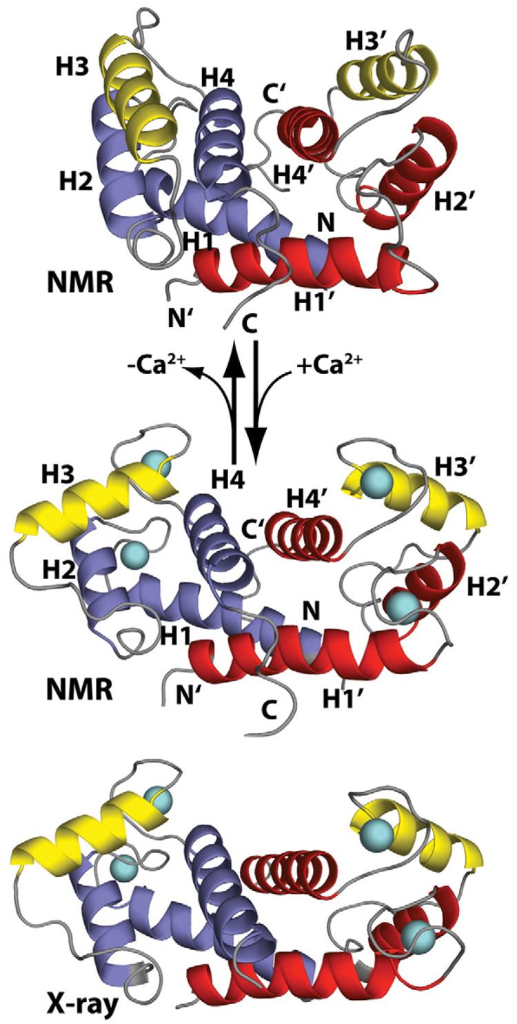
The “calcium switch” in S100B. Shown are ribbon diagrams of apo–S100B (NMR, PDB code: 1B4C) and Ca2+-bound S100B (NMR, PDB code: 1QLK; X-ray, PDB code: 1MHO) illustrating the calcium-dependent reorientation of helix 3 (yellow) in each S100B subunit, termed the “Ca2+ switch.” The other three helices in each subunit of the symmetric S100B homodimer are colored red and blue, respectively. The loop regions of S100B are colored gray, and the two calcium ions per subunit are cyan spheres. This calcium-dependent conformational change is required for S100B to interact with specific protein targets such as p53.