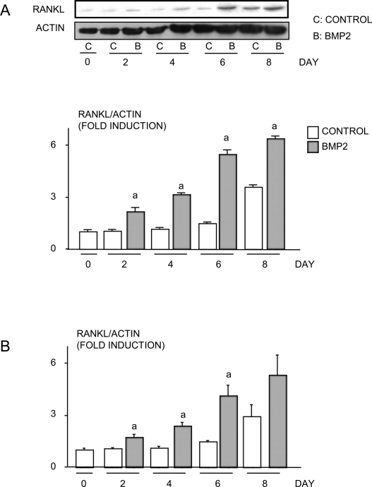
FIG. 2.
BMP2 increases RANKL protein expression in chondrocytes. (A) ATDC5 cells or (B) primary mouse sternal chondrocytes were treated with or without BMP2 (100 ng/ml) for various times. RANKL and β-actin protein levels were examined by Western blot analysis. A representative Western blot from ATDC5 cells is shown (top panels, A). Quantitation of protein bands was performed by densitometry using NIH image. The relative expression levels of RANKL protein were normalized by β-actin protein levels. The fold induction was measured by the following: the intensity of relative RANKL protein expression at each time-point/the intensity of relative RANKL protein expression in the day 0 sample. a p < 0.05 vs. control value at same time-point.

