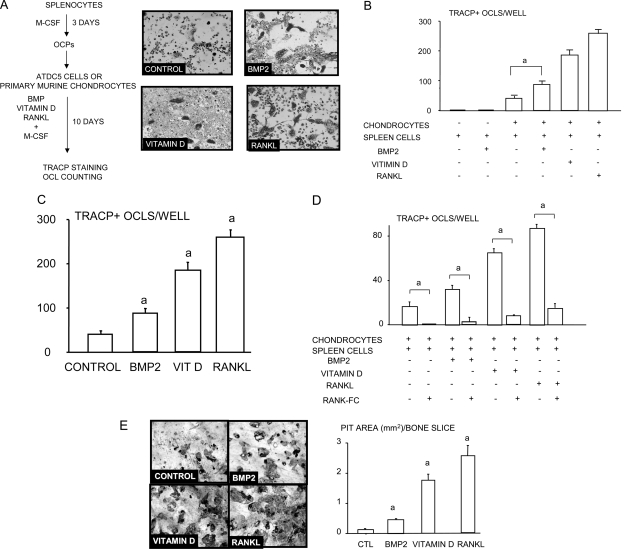
FIG. 3.
BMP2-stimulated chondrocytes induce osteoclast formation in vitro. ATDC5 cells were plated in subconfluent condition and were treated with or without BMP2 for 12 h. Spleen cells were cultured with conditioned medium containing M-CSF (1:20) for 3 days to generate osteoclast precursors. Osteoclast precursors were seeded on top of the chondrocytes. Co-cultures were maintained for an additional 10 days in the presence of BMP2, vitamin D3, or RANKL. Osteoclasts were detected by TRACP staining. Representative pictures from the co-cultures (A). The number of osteoclasts in BMP2-, vitamin D3– or RANKL-treated groups (B) and in RANK:Fc-treated groups (C). Co-cultures of chondrocytes and splenocytes were performed on bone slices for 14 days. After cells were brushed off, dentine slices were stained with 0.1% toluidine blue, and the total area of the pits per slice was measured (D). The same experiments were repeated twice with similar results. a p < 0.05 where indicated in B and C and vs. the control value in D.

