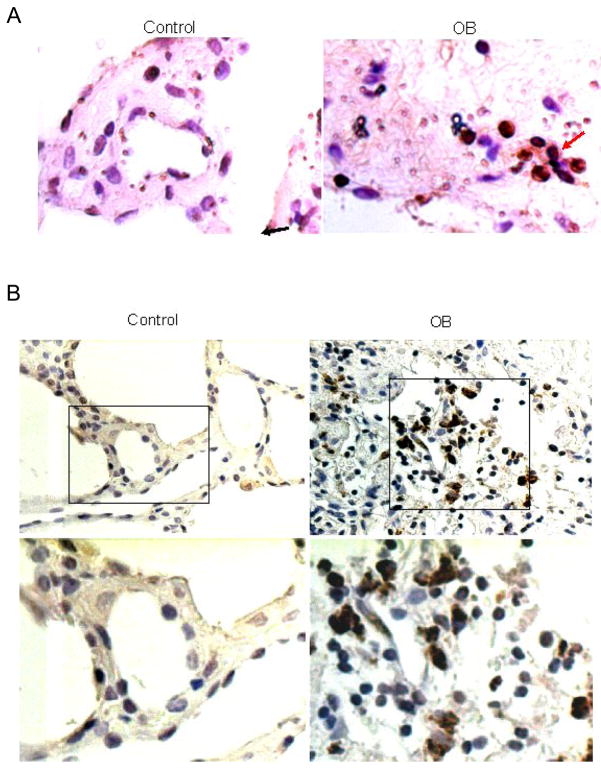
Figure 5.
Increased SDF-1 and CXCR-4 in OB lung samples compared to normal human lungs. Lung tissues from OB patients (right) and normal control (left) were paraffin embedded and sectioned. Immunohistochemistry was performed for SDF-1 (A) and CXCR-4 (B). Cells stained dark brown were SDF-1 or CXCX4 expressing cells (see arrows).

