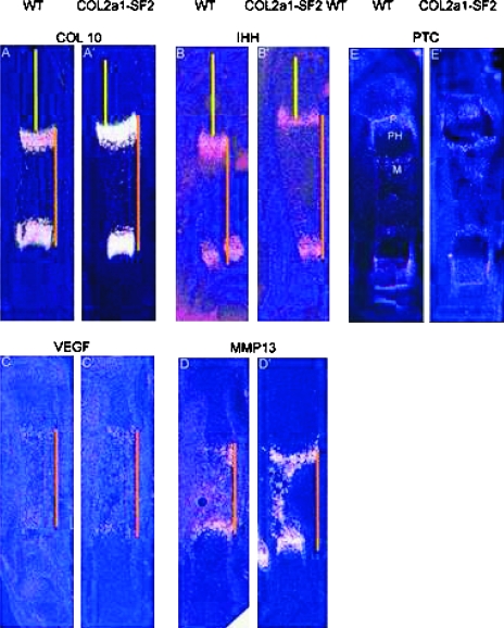
FIG. 3.
Advanced chondrocyte maturation in Col2a1-Smurf2 embryos. (A–D′) Chondrocyte maturation markers were analyzed on longitudinal sections through the tibia from wildtype (A–D) and Col2a1-Smurf2 (A′–D′) embryos by in situ hybridization. The orange line indicates the distance between the separated domains of Col10 (A and A′), Ihh (B and B′), VEGF (C and C′), and MMP13 (D and D′), and the yellow line indicates that from the domain to the articular surface. (E and E′) Ptc expression was analyzed on sections from wildtype (E) and Col2a1-Smurf2 (E′) by in situ hybridization. P, proliferation zone; PH, prehypertrophic zone; M, marrow cavity.

