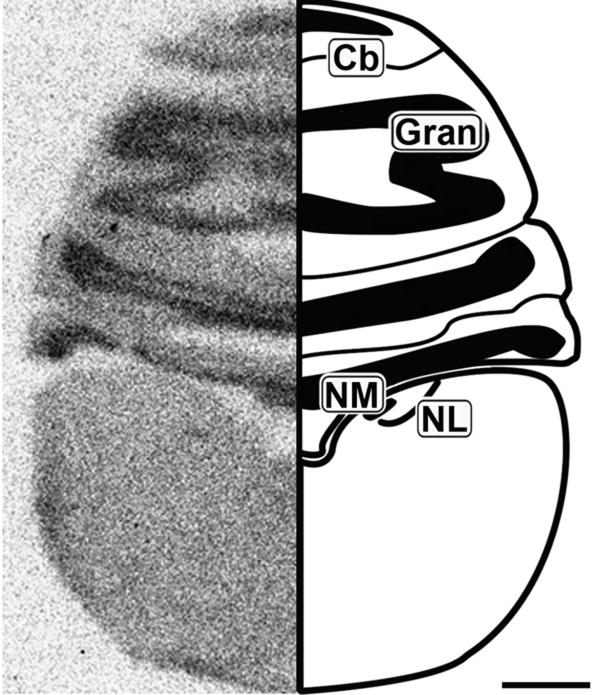
Figure 2.
Caudal coronal 20μm autoradiograph showing CB1 mRNA expression (left) and corresponding anatomical schematic (right). High levels of expression are observed in the granule cell region of the cerebellum. Cb: Cerebellum, Gran: Granule Cell Layer, NM: Nucleus Magnocellularis, NL: Nucleus Laminaris. Scale bar = 1mm.

