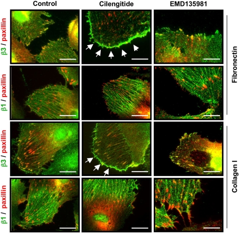
Figure 1. Cilengitide causes loss of αVβ3 from focal adhesions and promotes appearance of αVβ3 patches at the cell edge.
HUVEC were plated on coverslips coated with fibronectin or collagen I and were treated with 10 µM of cilengitide for 20 minutes. The localization of the αVβ3 or β1 integrin (green) and paxillin (red) were monitored by immunofluorescence staining. In HUVEC plated on fibronectin αVβ3 was present at focal adhesions, while β1 was present at fibrillar adhesions. Cilengitide, but not EMD 135981, caused loss of αVβ3 from focal adhesions and appearance of αVβ3-positive thin patches at the cell edge (arrows). β1 localization was not altered by cilengitide. A similar effect on αVβ3 (arrows) was observed on cells plated on collagen I, with the difference that focal adhesions were less abundant on this matrix. Optical magnification: 400×; Bar: 10 µm. (n = 5).

