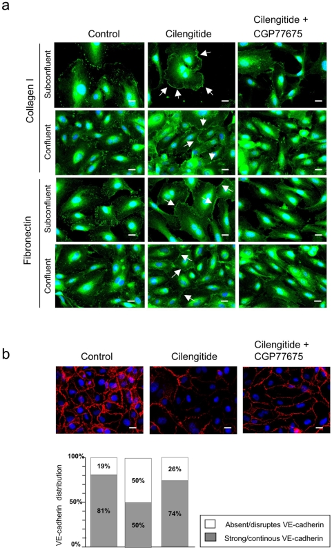
Figure 6. Src inhibition prevents cilengitide-induced relocalization of αVβ3 at the cell edge and disappearance of VE-cadherin from cellular junctions.
(a) Subconfluent and confluent HUVEC cultured on fibronectin or collagen I were exposed for 20 minutes to cilengitide (10 µM) in the presence of absence of CGP77675 (2.5 µM) and stained for αVβ3. Cilengitide-induced recruitment of αVβ3 to the cell edge (arrows) and this was prevented by CGP77675. (b) Confluent HUVEC cultured on fibronectin were treated for 20 minutes with cilengitide in the presence or absence of CGP77675. CGP77675 prevented cilengitide-induced VE-cadherin loss from cell-cell contacts. The bar graph gives the quantification of VE-cadherin staining at cell borders. The white and gray segments of the bars represent absent/disrupted vs. strong/continuous VE-cadherin staining, respectively (see material and methods for details). (n = 3). Optical magnification: 400×; Bars: 10 µM.

