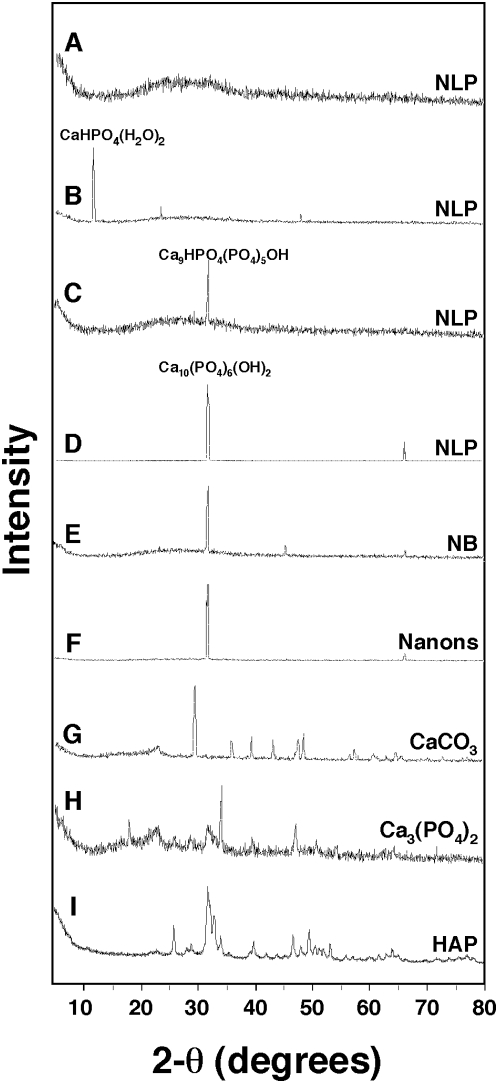
Figure 3. Powder X-ray diffraction analysis of NLP and NB.
(A–D) Various amorphous and crystalline patterns are associated with NLP. NLP were prepared from the inoculation of 3 mM each of CaCl2, Na2CO3, and NaH2PO4 into DMEM. NLP were examined by XRD (A,B) immediately after formation, which revealed either an amorphous pattern (A) or the presence of the mono-calcium form CaHPO4(H2O)2, or they were examined after the following periods of incubation in DMEM at 37°C: (C) 1 day; and (D) 4 days. (E) NB obtained from DMEM containing 10% HS. (F) “Nanons” after one week in DMEM without serum. Peaks were assigned the specified chemical structure after comparing peaks with the database. With the exception of (C), which revealed the Ca9 form of HAP, the peaks seen in (D–F) all corresponded to Ca10 HAP. Peaks at 32.0 degrees and 31.8 degrees correspond to the Ca9 and Ca10 forms of HAP, respectively. Notice also the small peaks at 66.2 degrees seen only with the Ca10 form (D–F). Commercial powders of (G) CaCO3, (H) Ca3(PO4)2, and (I) HAP were included as controls.

