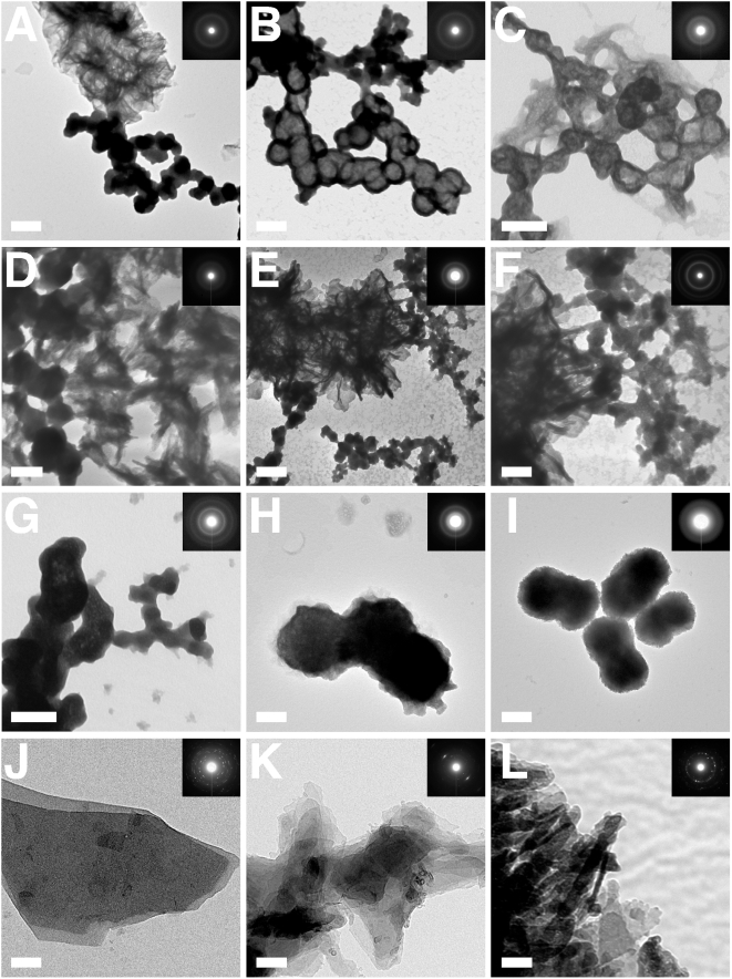
Figure 6. Negative-staining TEM and electron diffraction patterns of NLP and NB.
(A–G) NLP were prepared as in Fig. 3 and submitted to TEM after incubation in DMEM at 37°C for either 2 days (A–C, G), 2 weeks (D), or 1 month (E and F). Round, dividing, or budding NLP are shown in close association with a crystalline phase, which shows either as a network of filamentous/membranous materials or as spindles/needles. The electron diffraction pattern characteristic of polycrystalline material obtained for both phases is shown in the inset. (B–D) show NLP with coccoid shapes and diameters smaller than 100 nm among larger particles. (D–F) Crystalline biofilms associated with NLP are seen with longer incubations. (F) is a magnified image of (E) depicting the transition between the round NLP and the crystalline matrix. (H and I) NB showing cell-dividing forms similar to NLP. (H) NB obtained from HS and (I) “nanons,” as in Fig. 3. Small crystalline projections can be distinguished on their surface. NB look virtually indistinguishable from NLP (compare with image G taken of NLP). Commercial (J) CaCO3, (K) Ca3(PO4)2, and (L) HAP produce a higher degree of crystallinity compared to NLP, NB, and “nanons” (insets). Scale bars: 100 nm (B–D, H–J, L); 200 nm (A, E–G, K).

