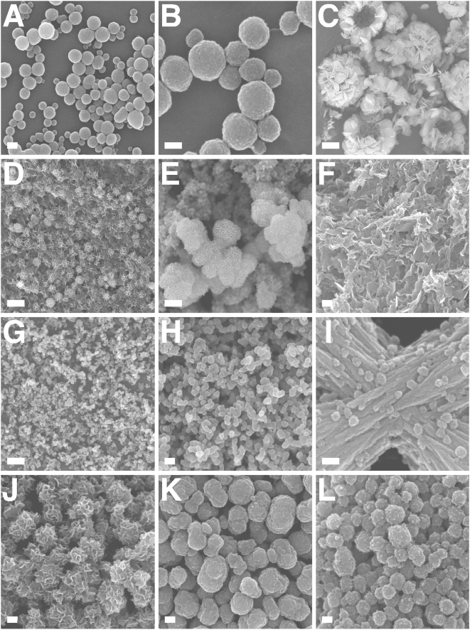
Figure 7. Identical morphologies of NLP and NB seen by field-emission SEM.
(A–F) NLP were obtained as in Fig. 3 and incubated in DMEM for either 2 days (A and B), 4 days (D and E) or 1 month (C and F). (A) NLP consisted of spherical particles with a few dumbbell formations suggestive of cell division. (B) Enlarged image of NLP presented in (A) showing round particles with a size ranging 50–200 nm. (C) Conversion of round NLP to crystalline HAP in the form of needle-like formations along with large exposed hollow spheres in the shape of “igloos” and “shelter” forms of NB (see ref. 4). (D) Conversion of NLP into a crystalline film-like matrix. (E) Enlarged image of (D), showing crystalline surface of NLP, with their rough surface coated with small needle-like crystals. (F) Dense crystalline matrix formed after prolonged incubation of NLP. (G) Small-sized NLP obtained in the presence of excess magnesium and carbonate. NLP were prepared from the inoculation of 3 mM CaCl2, 18 mM Na2CO3, 3 mM NaH2PO4, and 3 mM MgCl2 into DMEM. (H) Enlarged image of NLP seen in (G), showing sizes typically around 100 nm. (I) NLP prepared as in (A), showing round particles attached to filamentous and membranous material. (J–L) NB with morphologies similar to those of NLP. (J) NB cultured from HS, as in Fig. 6, consisting of coalesced particles covered with needle-like formations. (K) “Nanons” and (L) NB strain DSM 5819 cultured in DMEM for one week without serum, showing round, ellipsoid and division-like large formations. (L) NB strain DSM 5819. Scale bars: 100 nm (B, I); 200 nm (A, C, F, H, J–L); 1 µm (G); 2 µm (C); 5 µm (D).

