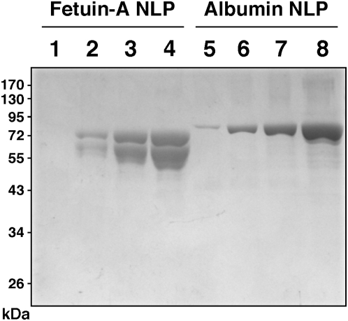
Figure 21. Demonstration by SDS-PAGE of the presence of fetuin-A and albumin in NLP formed from inhibitory complexes.
NLP were prepared as in Figs. 19 and 20, from 3 mM each of CaCl2, Na2CO3, and NaH2PO4 in 1 ml of water incubated with either BSF or HSA at the following concentrations: 40 µg/ml, 80 µg/ml, 160 µg/ml, and 320 µg/ml of BSF, corresponding to lanes 1–4, respectively; and 0.2 mg/ml, 0.4 mg/ml, 0.8 mg/ml, and 1.6 mg/ml of HSA, for lanes 5–8, respectively. After an incubation of 3 days at room temperature, centrifuged pellets were washed three times with HEPES buffer, and the final washed pellets were resuspended in 50 µl of double-distilled water containing 50 mM EDTA, of which 16 µl were loaded onto each lane. Note the presence of multiple bands around 50–70 kDA associated with BSF and a prominent band centered around 72 kDa corresponding to HSA.

