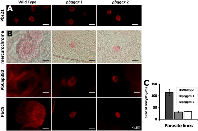
Figure 5. Development of pbggcs− oocysts in A. stephensi mosquitoes.
(A) Oocysts on dissected midguts at 2 d.p.i., reacted with antibody Mab 13.1 against the ookinete/young oocysts surface protein Pbs21. Oocysts were detected after incubation with a Rhodamine Red™-X goat anti-mouse IgG (H+L) secondary antibody (Invitrogen). No differences in the reactivity pattern of oocysts from pbgccs − and wild type parasites were observed. (B) Oocysts on dissected midguts at day 12 p.i. stained with 0.2% mercurochrome (upper panel), antibodies against PbCap380 in the oocyst capsule (center panel) and antibodies against the surface protein of sporozoites PbCS (Mab 3D11). Antibody-stained oocysts were detected by indirect fluorescence microscopy using Rhodamine Red™-X goat anti-mouse IgG (H+L) or Texas Red®-X goat anti-rabbit IgG (H+L) secondary antibodies (Invitrogen), respectively. The pbggcs− oocysts produced the PbCap380 protein but are strongly reduced in size compared to wild type (see also C). Anti-CS staining shows that, although CS is produced, 12 day old oocysts lack the typical CS staining pattern of wild type oocysts. (C) Oocyst size on dissected midguts 12 d.p.i.

