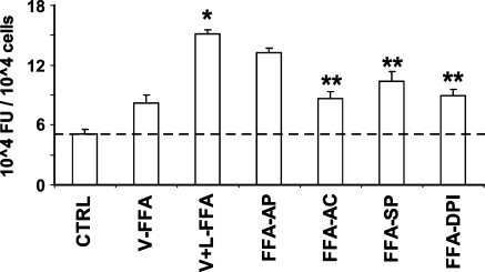
Fig. 5.
Effects of FFA isolated from VLDL (V-FFA) and VLDL lipolysis (V + L-FFA) on ROS production in HAECs in the absence or presence of enzyme inhibitors. Each lipid fraction was isolated from VLDL (50 mg/dl TG), evaporated, redissolved, and delivered to cells in a PBS vehicle (5 μl). HAECs were preincubated for 30 min with the fluorescence probe DCF-AM (10 μM), followed by incubation for 2 h with FFA isolated from VLDL with or without 30 min incubation with LpL (2 U/ml) in the absence or presence of allopurinol (FFA-AP) (100 μM), apocynin (FFA-AC) (100 μM), sulfaphenazole (FFA-SP) (10 μM), or DPI (FFA-DPI) (50 μM). Fluorescence intensity of cells was measured with a fluorescence microplate reader. Fluorescence distribution of DCF-AM oxidation was expressed as fluorescence units (FU) and expressed as the mean ± SEM (n = 6). Significant differences in means were determined by 2-tailed Student's t-test (* P < 0.05 vs. V-FFA; **P < 0.05 vs. V+L-FFA).

