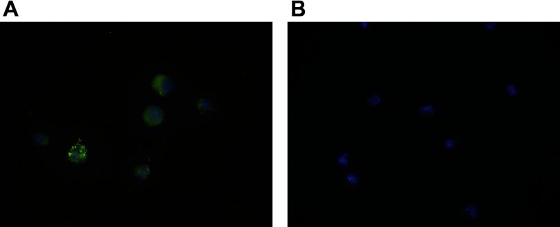
Fig. 7.
Immunofluorescent staining of LBP on C57BL/6 Kupffer cells. Four days following CBDL, Kupffer cells isolated from C57BL/6 mice demonstrate the LBP is present on the cell surface as well within the cell (A), as demonstrated by the confocal microscope images, ×100 magnification. The LBP appears green (stained with Alexa Fluor 488), whereas the nuclei appear blue (stained with DAPI). Kupffer cells isolated from LBPKO CBDL mice (negative control) demonstrate only positive nuclear staining (B).

