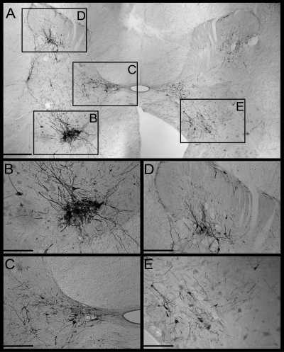
Fig. 1.
Photomicrographs of infected neurons in the C5 spinal cord of animal C21. A: photomontage of the entire C5 spinal gray matter of one section. Boxes denote regions depicted at higher magnification in subsequent panels. The left side of the section is ipsilateral to the rabies injections into the diaphragm. B: a dense cluster of presumed motoneurons in the ventral horn. C: a group of infected presumed interneurons in Rexed's lamina X and the medial portion of lamina VII. D: a group of infected presumed interneurons concentrated in Rexed's lamina V. E: infected presumed interneurons in medial lamina VII and lamina VIII contralateral to the side of injections. Bars designate 500 μm in A, and 250 μm in the other panels.

