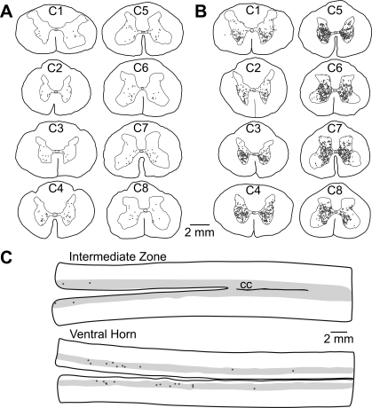
Fig. 2.
Locations of presumed interneurons labeled following the injection of rabies virus into the diaphragm. A and B: locations of labeled cervical interneurons in an early infection (animal C21; A) and an intermediate infection (animal C51; B) case. Representative transverse sections from each cervical segment are shown; the level is indicated on the section. C: locations of labeled interneurons in horizontal sections through the T5–T9 thoracic spinal cord of intermediate infection case C38. The gray matter is indicated as a shaded area, and the rostral end of the sections is toward the left side. The top section is through the intermediate zone, the portions of Rexed's lamina VII and X near the level of the central canal (CC). The bottom section is through the ventral horn. No sections that were clearly through the dorsal horn contained any infected neurons and thus are not provided.

