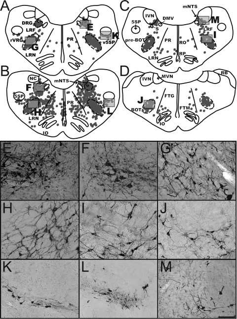
Fig. 3.
Distribution of labeled neurons in the medullary respiratory groups and adjacent regions. A and B: maps of sections from early infection (animal C21; A) and intermediate infection (animal C39; B) cases illustrating the localization of rabies-infected neurons within the dorsal respiratory group (DRG) and rostral ventral respiratory group (rVRG) at 12.1 mm posterior to the interaural plane. C: map of a section through the pre-Bötzinger (pre-BOT) complex in early infection case C21 at 11.6 mm posterior to the interaural plane. D: map of a section through the Bötzinger (BOT) complex in early infection animal C52 at 10 mm posterior to the interaural plane. Each dot in the maps represents a single labeled neuron. Boxes indicate the areas depicted in the photomicrographs at the bottom of the figure. E and F show the DRG, G and H the rVRG, I the pre-BOT, J the BOT, K and L the ventral paratrigeminal nucleus (v5SP), and M the region of nucleus tractus solitarius (NTS) adjacent to the inferior vestibular nucleus (IVN). Most of the micrographs are from the same sections used to generate the maps, although F and H are from the intermediate infection case C51 (where the contrast between the labeled neurons and background was clearer than in animal C39). Scale bars in each photomicrograph represent 250 μm. 5SP, spinal trigeminal nucleus; DMV, dorsal motor nucleus of the vagus; FTG, gigantocellular tegmental field; FTM, magnocellular tegmental field; IO, inferior olivary complex; LRF, lateral reticular field; LRN, lateral reticular nucleus; mNTS, medial nucleus of the solitary tract; MVN, medial vestibular nucleus; NC, cuneate nucleus; PR, paramedian reticular nucleus; RB, restiform body; RO, raphe obscurus; RP, raphe pallidus; S, solitary tract.

