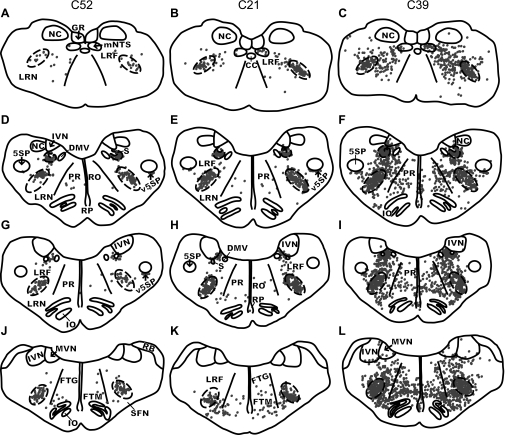
Fig. 4.
Maps of sections through the caudal and intermediate regions of the medulla in two early infection (C52, left column; C21, middle column) cases and one intermediate infection (C39, right column) case. Each dot represents a single infected neuron. The DRG and VRG respiratory columns are depicted as dashed areas on each map. A–C: the medulla at the level of the caudal VRG at 16 mm posterior from the interaural plane. D–F: the DRG and the rostral VRG at 12.1 mm posterior from the interaural plane. G–I: the pre-BOT complex at 11.6 mm posterior from the interaural plane. J–L: the BOT complex at 10 mm posterior from the interaural plane. Because of slight differences in the plane of cutting, sections at the same approximate anteroposterior level had different shapes and contained different structures. GR, gracile nucleus.

