Fig. 7.
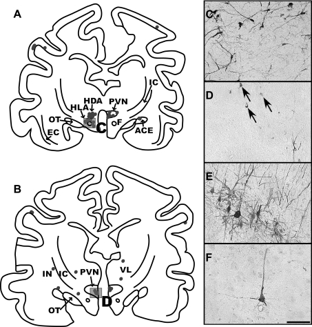
A and B: maps of sections through the diencephalon of intermediate infection animal C51. The section shown in A is located at 11 mm anterior to the interaural plane, and B is at 11.8 mm anterior to the interaural plane. Due to the plane of sectioning, the dorsal portion of sections is more rostral than the ventral part, and the right side of sections is more rostral than the left side. This is especially evident in A, where the left side contains labeled neurons in the dorsal (HDA) and lateral hypothalamic areas (HLA), consistent with infection of the perifornical region; in contrast, the right side of the section shows infection in the caudal extent of the paraventricular nucleus of the hypothalamus (PVN). The photomicrograph in C shows the morphology of labeled neurons in the perifornical area of the hypothalamus of a late infection case, animal C36; a box in A indicates the area that is depicted. The photomicrograph in D is from animal C51 and illustrates the morphology of infected neurons in the parvocellular division of the PVN, which are demarked by arrows; a box in B indicates the region illustrated. E and F: photomicrographs of infected neurons in the precruciate gyrus of a late infection (C36) and an intermediate infection (C39) animal, respectively. The cortex from both of these cases was cut in the sagittal plane. The scale bar represents 250 μm. ACE, central nucleus of the amygdala; EC, external capsule; F, fornix; IC, internal capsule; IN, insular cortex; OT, optic tract; VL, ventrolateral nucleus of the thalamus.

