Fig. 3.
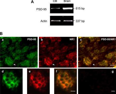
A: RT-PCR demonstrating message for PSD-95 in carotid body (CB, left lane) as well as in brain cortex (Brain, right lane). B: immunofluorescent staining showing colocalization of PSD-95 and NMDAR1 in glomus cells of the carotid body. a: Intense immunoreactive products for PSD-95 (green). b: Immunoreactive products for NMDAR1 (NR1, red). c: Merged images of a and b. d, e, and f: Higher magnification of the framed areas in a, b, and c, respectively. g: Negative control obtained by omitting the primary antibody in a consecutive section of c in Fig. 1B. The magnifications of a–c and d–f are same. Scale bars = 20 μm.

