Fig. 4.
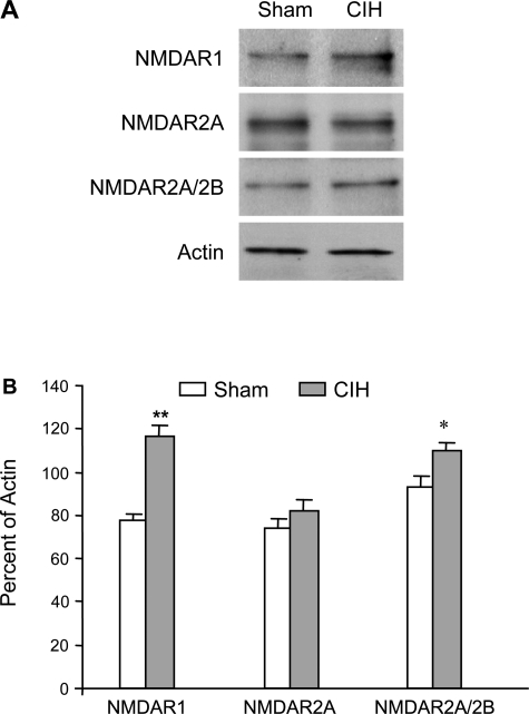
Western blot showing enhanced expression of protein products of NMDAR1 in carotid bodies of cyclic intermittent hypoxia (CIH)-exposed rats relative to sham-exposed animals. Homogenized tissues of carotid bodies pooled from 8 rats in each group were used. A: representative Western blots. B: percentages of NMDAR1, NMDAR2A, and NMDAR2A/2B to actin. Data (means ± SD of densitometric analysis) are from 3 individual Western blots run in triplicate. *P < 0.005, **P < 0.01 vs. sham rats.

