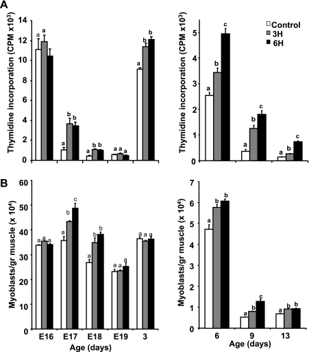
Fig. 3.
Thymidine incorporation by DNA (A) and number of muscle cells per gram of pectoralis muscle (B) in control and TM (3H, 6H) groups. Muscle was removed from embryos or posthatch chicks on various days and pooled within each group. Left: embryonic days (E) 16–19 and posthatch day 3; right: posthatch days 6–13. Myoblasts were isolated in parallel from each group and counted using a hemocytometer. Cells were incubated for 1 day, after which labeled thymidine was added for 2 h. Results are means ± SE (n = 6) of a representative of three independent experiments. a,b,cData with different letters differ significantly within the same age group (P < 0.05).

