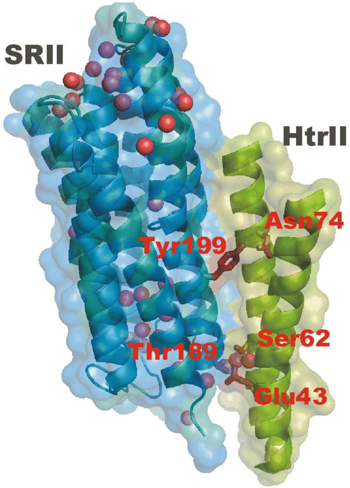
Figure 1.

An X-ray crystallographic structure of the SRII-HtrII complex (Protein Data Bank (PDB) access code 1H2S) with the focus on the receptor-transducer interface region with intermolecular Tyr199-Asn74 and Thr189-Glu43/Ser62 hydrogen bonds shown. Internal water molecules are shown as spheres. The figure was prepared using the program PyMOL (http://www.pymol.org).
