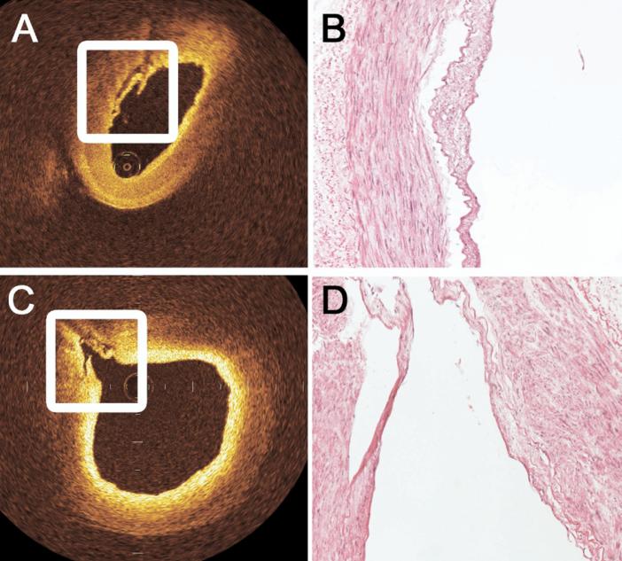
Fig 1.
Histologic confirmation of intimal injury was noted by the ex vivo optical coherence tomography (OCT) examination on (A) the luminal surface of the radial artery and (C) within the ostia of branch points exceeding 0.3 mm in diameter with thickened intima near the ostium. Image-guided biopsy specimens were obtained from areas suggested to be abnormal by OCT. As shown by these representative examples of (B) severe and (D) minor injury, registered histologic sections confirmed the diagnosis of intimal injury on every occasion and illustrate the near histologic resolution of OCT. (A: original magnification ×20, eosin stain; B: original magnification ×20, eosin stain.)

