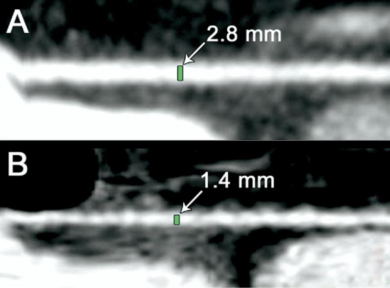
Fig 3.
Postoperative evaluation of radial artery (RA) spasm using 64-multislice detector computed tomography. Two-dimensional planar image reconstructions were obtained parallel to the long axis of the RA to directly measure the diameters of the bypass grafts. These representative examples of RA grafts were imaged on postoperative day 5 after coronary artery bypass grafting. (A) Patient with a RA graft with an unaffected diameter. (B) Radial artery graft with “string sign.”

