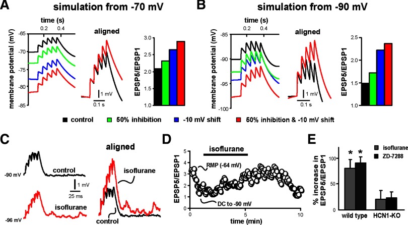
FIG. 6.
Isoflurane enhances excitatory postsynaptic potential (EPSP) temporal summation in cortical pyramidal neurons from WT mice, but not from HCN1-KO mice. A and B: computer simulations in a cortical neuron model from 2 different initial potentials (−70 and −90 mV) illustrate effects on EPSP summation accompanying a 50% decrease in Ih amplitude, −10-mV shift in V1/2 of activation, or both. Aligned traces illustrate the enhanced summation observed in the model with both a decrease in Ih amplitude and a shift in V1/2, and histogram plots show effects on EPSP summation of independently or coordinately changing these 2 parameters in the model. C: sample voltage traces show EPSP recordings obtained from cortical pyramidal neuron in response to 40-Hz, 7-V stimulation under control conditions and after incubation in 0.4 mM isoflurane (right). To enhance basal dendritic Ih, current injection was used to produce an initial somatic membrane potential of −90 mV; EPSPs are aligned to initial membrane potential and normalized to the amplitude of the first EPSP in the train to highlight enhanced temporal summation of EPSPs. D: summation was quantified as the amplitude ratio of the 5th to 1st EPSP and plotted with respect to time; isoflurane enhanced temporal summation. Note that DC injection to hyperpolarize the neuron caused a decrease of EPSP summation, consistent with a contribution of Ih to the sublinear summation under control conditions. E: averaged percentage increase of EPSP summation (ratio of EPSP5/EPSP1, relative to control) induced by 0.4 mM isoflurane or 50 μM ZD-7288 or in WT and HCN1-KO mice; isoflurane and ZD-7288 enhanced EPSP summation in cortical neurons from WT animals (*P < 0.05 vs. control), but had no effect in cells from HCN1-KO mice.

