Figure 7.
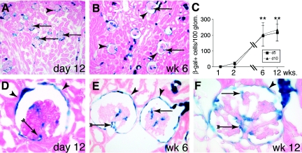
Recruitment of PECs onto the glomerular tuft in adolescent pPEC-rtTA/LC1/R26R mice. (A) After genetic tagging of PECs 5 d after birth, β-gal–positive cells (arrows) can be detected on glomerular tufts on day 12. (B) Six weeks after birth, genetically tagged cells are present within most glomeruli (arrows) of the outer cortex as well as close to the medulla. Genetic labeling persists in PECs (arrowheads). (C) Statistical analysis of β-gal–positive cells per 100 glomeruli over time in triple-transgenic PEC-TETon mice induced 5 (d5) or 10 d after birth (d10). A similar increase of β-gal–positive cells over time was observed in both groups (**P < 0.01 ANOVA; n = 5 for each time point). (D through F) Genetic labeling persists in PECs (arrowheads) 12 d (D) and 6 and 12 wk (E and F) after doxycycline administration. β-Gal–positive cells were identified close to the VP (arrow with tails) as well as projecting into the periphery of the glomerulus (arrow). (F) Occasionally, glomeruli with up to 20 β-gal–positive cells were observed at 12 wk of age (arrowheads, labeled PECs; A, B, and D through F, X-gal/eosin staining on 6-μm cryosections).

