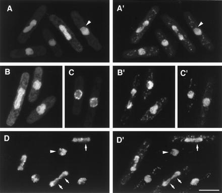
Figure 2.

GFP fusion proteins localized to the nucleus. (A–D) GFP fluorescence; (A′–D′) corresponding images of DNA staining by PI. (A and A′) Localization of M31. (B and B′) Localization of M30 at low expression levels. (C and C′) Nuclear “rim” localization of M30 at higher expression levels (see text). (D and D′) Localization of M25 to nuclear chromatin. Note lack of staining in the interphase nucleolus, which stains weakly with PI (arrowhead), and lack of association of M25 with specific strands of segregating chromatin (arrows), which may be embedded in nucleolar material. (Bar = 5 μm.)
