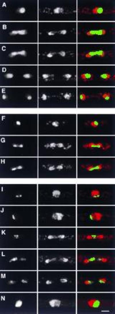
Figure 3.

GFP fusion proteins with nucleolar localizations. (Left) GFP; (Center) DNA (PI stain); (Right) merged image of GFP (green) and DNA (red). (A–E) General nucleolar localization of S32, during interphase (A) and different stages of mitosis (B–E). (F–H) Localization of S2 in interphase (F) and mitosis (G and H). Note the more compact organization of S2 in mitosis relative to S32. (I–N) Punctate nucleolar localization of S19 in interphase (I and J) and mitosis (K–M), and more general nucleolar localization when highly overexpressed (N). Note the specific association of S19 and trailing chromatin strands in L. (Bar = 2 μm.)
