Figure 4.

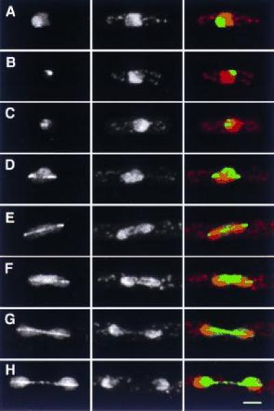
Localization of S26 to the nucleolus during interphase, and to the nucleolus and mitotic spindle during mitosis. (Left) GFP; (Center) DNA (PI stain); (Right) merged image of GFP (green) and DNA (red). (A–C) Interphase cells. Note heterogeneity of fluorescence, which in all cases is confined to the nucleolus. (D–H) Different stages of mitosis. During mitosis, S26 remains associated with nucleolar regions, albeit to varying degrees in different cells (compare E with F, and G with H). (Bar = 2 μm.)
