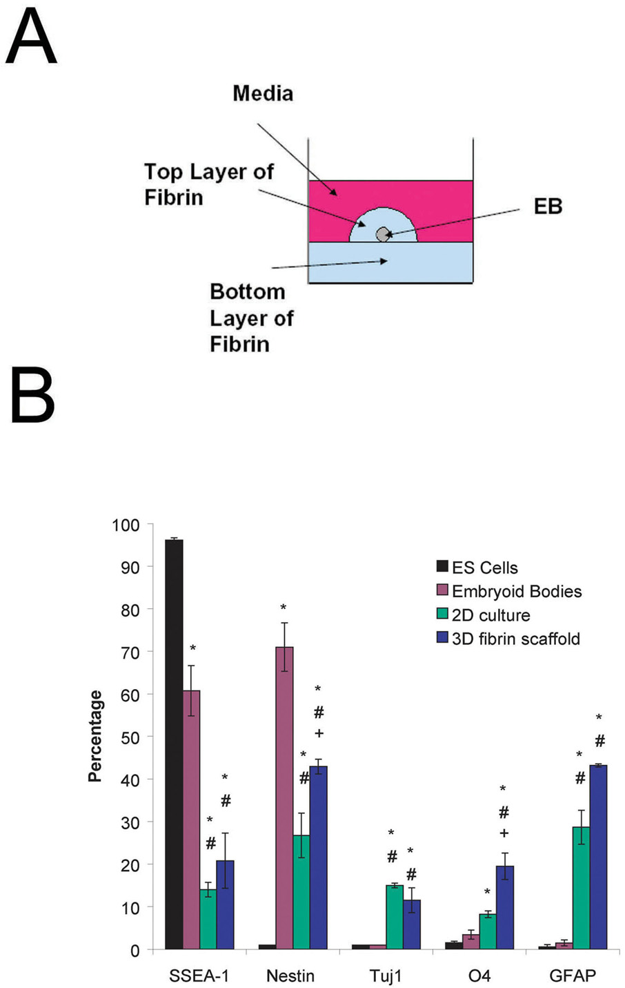
Figure 1. Schematic of 3 dimensional culture system and results of preliminary characterization of ES cell cultures using FACS.
A) Side view of an individual well of a 24 well plate showing the placement of the EB and surrounding fibrin scaffold. B) Results of FACS analysis performed on undifferentiated mouse ES cells, 4−/4+ embryoid bodies, 2D culture and EBs seeded into fibrin scaffolds after 14 d of culture when no growth factors are present. The markers examined are as follows with the phenotype description given in parentheses: SSEA-1 (undifferentiated mouse ES cells), nestin (neural precursors), Tuj1 (neurons), O4 (oligodendrocytes), and GFAP (astrocytes). * indicates p < 0.05 for that marker compared to undifferentiated ES cells. # indicates p <0.05 for that marker compared to embryoid bodies. + indicates p < 0.05 for that marker compared to the 2D culture.

