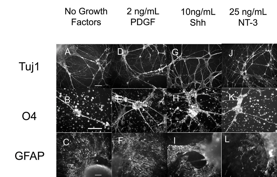
Figure 2. Immunohistochemistry performed on EBs after 14 days inside of fibrin scaffolds for mature cell markers including Tuj1 (neurons), O4 (oligodendrocytes) and GFAP (astrocytes).
A–C) In scaffold staining of EB culture when no growth factors were present. D–F) In scaffold staining of EB culture when 2 ng/mL of PDGF was present. G–I) In scaffold staining of EB culture when 10 ng/mL of Shh was present. J–L) In scaffold staining when 25 ng/mL of NT-3 was present. Scale bar for Tuj staining are 100 µm. Scale bars for O4 and GFAP staining is 50 µm.

