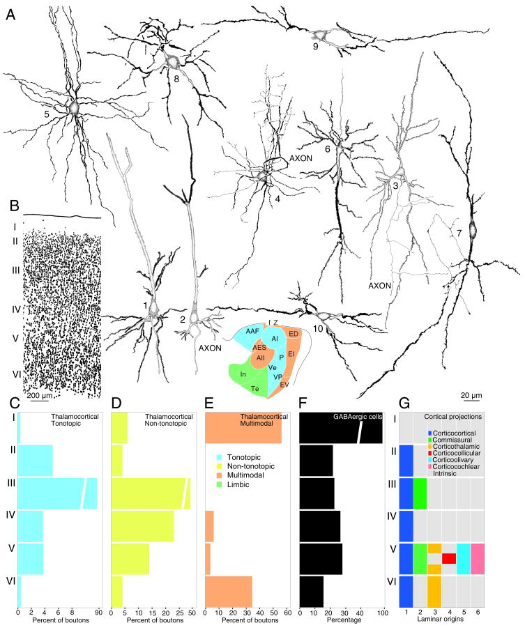
Fig. 1.
Some basic anatomical features of cat auditory cortex (AC) area AI (primary AC). (A) Major cell types include glutamatergic pyramidal cells (1–3), GABAergic basket (4) and multipolar (5) neurons, spinous inverted pyramidal cells (6), bipolar cells (7), small multipolar neurons (8), and horizontal cells in layers I (9) and VI (10). Many more subtypes are found in morphological studies limited largely to AI (Winer, 1992). Golgi-Cox impregnation, 140 μm thick section, planachromat, N.A. 0.95, ×1000. (B) AI cytoarchitecture shows a prominent layer I, a dense concentration of layer II cells, smaller layer IV cells, and columnar somatic arrangements in deeper layers. Nissl preparation, 30 μm thick celloidin embedded section, planapochromat, N.A. 0.65, ×500. (C–E) Laminar distribution of thalamocortical boutons after medial geniculate body (MGB) deposits of biotinylated dextran amines (Huang and Winer, 2000). Abscissa, bouton percentages/layer in pia-white matter traverses. (C) After ventral division tracer deposits, labeling is concentrated in layer III. (D) MGB dorsal division deposits had a wider laminar dispersion. (E) Medial division deposits had a bilaminar AC pattern. (F) The proportion of γ-aminobutyric acid-containing (GABAergic) AI cells is lamina specific. Antisera to GABA, 30 μm thick frozen section (Prieto et al., 1994b). (G) The laminar origins of six AI projection systems (1–6). While these origins overlap, few such neurons project to multiple subcortical targets (Wong and Kelly, 1981); gray background, intrinsic and local projections (Read et al., 2001), also present within the colored regions. 1: unpublished observations; 2: Code and Winer (1985); 3: Winer and Prieto (2001); 4: Winer (2005); 5: Schofield and Coomes (2004); 6: Schofield and Coomes (2005). For abbreviations, see the list.

