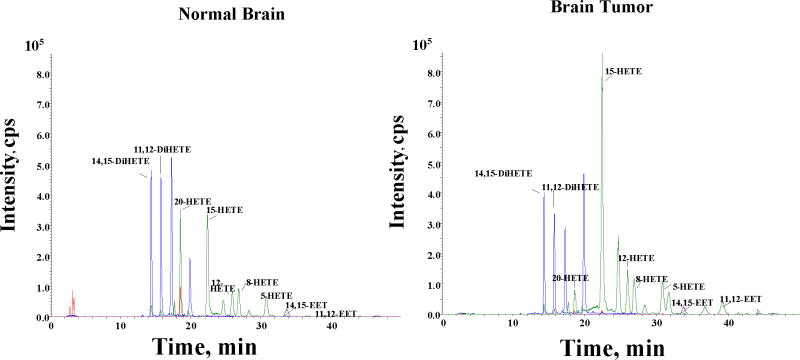
Figure 6.
Representative LC-MS profile of CYPAA metabolites in normal and tumor rat brain tissue samples. Normal brain and tumor core tissue were removed from rat brain and frozen in liquid nitrogen. The samples were homogenized and assayed for CYP activity as described in the methods section. Epoxyeicosatrienoic acids and Di-HETEs (8, 9- 11, 12- and 14,15-EETs) and HETEs ( 5-, 8-, 12-,15- and 20-HETE) were detected in both normal and tumor tissues.

