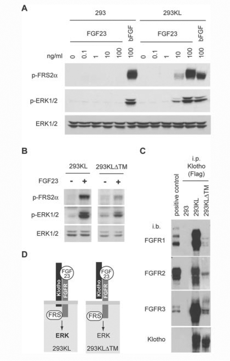
Figure 3. FGF23 requires Klotho to activate FGF signaling.
A, 293 cells or Klotho-expessing 293 cells (293KL) were stimulated with various concentration of recombinant human FGF23 or basic FGF (100 ng/ml) for 15 min. Activation of FGF signaling was determined by immunoblot analysis using anti-phospho-FRS2a antibody (p-FRS2a), anti-phospho-ERK1/2 antibody (p-ERK1/2), or anti-ERK1/2 antibody (ERK1/2). B, 293KL cells or 293KLDTM cells were stimulated with the conditioned medium containing mouse FGF23 (R179Q) (~10 ng/ml) for 15 min. The same volume of conditionedmedium from mock-transfected 293 cells was used as a negative control (FGF23−). C, detection of endogenous FGFRs bound to Klotho in 293KL cells. Lysates of 293, 293KL, and 293KLDTM cells were immunoprecipitated with Klotho using anti-FLAG antibody and then immunoblotted with antibodies against FGFR1, FGFR2, FGFR3, and Klotho. Lysates of 293 cells transfected with expression vectors for mouse FGFR1c, FGFR2c, and FGFR3c were immunoprecipitated with anti-V5 antibody and used as positive controls for immunoblotting of FGFR1, FGFR2, and FGFR3, respectively. D, a model for interaction between Klotho, FGFR, FGF23, and FGF signaling.

