
Figure 1.
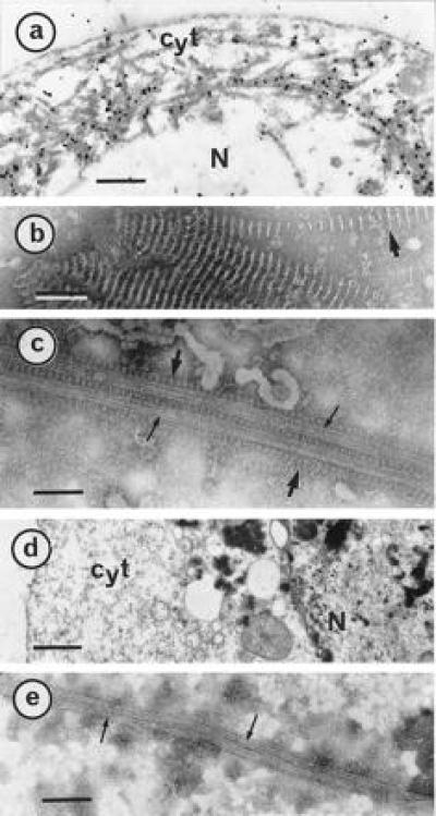
P18 paracrystals decorate MTs within Sf9 cells. Cells infected for 3 days were harvested and processed for electron microscopic observations and gold-labeling as described (15). (a) A thin section of an infected Sf9 cell was gold-labeled by a P18 antibody. As reference, negatively stained characteristic P18 paracrystals (15) are shown in b. (c) Infected cells were treated with Taxol and disrupted in AB buffer before negative staining. P18 paracrystals and well-shaped MTs are indicated with large and small arrows, respectively. (d) Thin section of a cell infected with a wild type baculovirus. (e) Negative staining of a similar cell disrupted after a Taxol treatment. Cyt, cytoplasm; N, nucleus. [Bars = 1 μm (a and d), 100 nm (b), and 75 nm (c and e).]