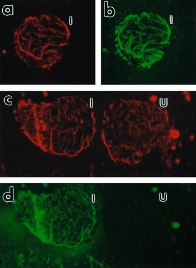
Figure 5.

Fluorescence micrographs of Nicotiana tabacum cv. Xanthi membrane ghosts. Membrane ghosts were double labeled as indicated for visualizing both MTs (a and c) and P18 (b and d). The same membrane ghost from an infected protoplast producing P18 is shown in a with a rhodamine filter cube for visualizing the MTs and in b with a fluorescein filter cube for visualizing P18. After Taxol treatment (see text), MTs of two membrane ghosts are shown as a rhodamine fluorescence in c and the presence of P18 in only one of these cells is shown by fluorescein in d. I, infected cell; U, uninfected cell. (×400 for a–d.)
