
Figure 7. Disruption of GIPC suggests that Glut1 can exist in a recycling pool distinct from TfR.
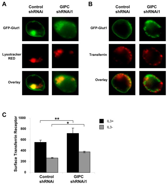
(A and B) FL5.12 cells were transfected with GFP-Glut1 and control shRNAi or GIPC shRNAi1 constructs for 48 hours and were incubated with (A) 50 nM Lysotracker RED for 30 minutes or (B) 50μg/ml Alexa Fluor 568-tagged Transferrin for one hour followed by fixation and visualization with fluorescence microscopy. (C) Cells were transfected with control shRNAi or GIPC shRNAi constructs for 48 hours and then were cultured in the presence or absence of IL3 for an additional six hours. Cells were stained with anti-TfR antibody and surface levels of TfR were measured via flow cytometry. Mean and standard deviations of triplicate samples are shown. Asterisks (*) indicate p< 0.0005 and (**) p≤ 0.05 within the experiment. Representative results are shown for three or more experiments.