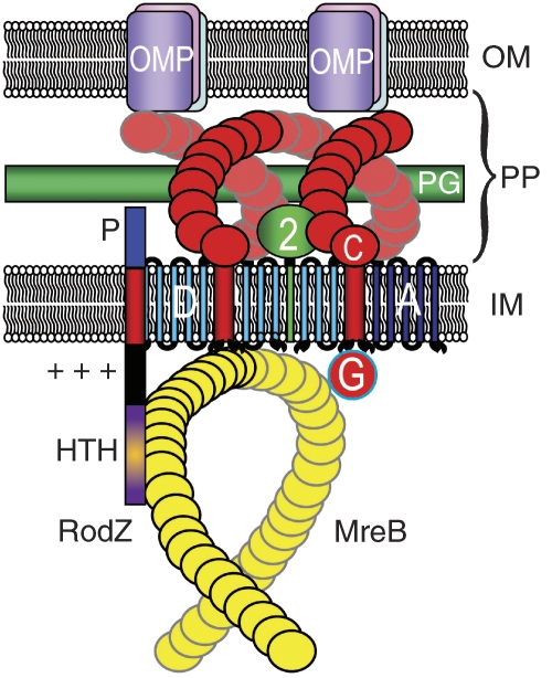
Figure 1.
Schematic diagram that visualizes the interactions between RodZ and the MreBCD–MrdAB (PBP2 RodA) complexes of E. coli. RodZ is shown as a vertical bar with four domains. The HTH domain (magenta) mediates interaction with MreB, whereas the juxta-membrane (JM) domain (+++) is in close contact with the negatively charged membrane phospholipids and serves to configure MreB in its helix-like appearance. P is the periplasmic part of RodZ that interacts with as yet unknown components in the periplasm. MreB is shown as a yellow helix beneath the inside of the inner membrane (IM) that interacts with MreC (red helix). MreC, in turn, interacts with PBP2 (green) and different OMPs. PP, periplasm; OM, outer membrane; PG, peptidoglycan layer; D, MreD in the IM; A, RodA in the IM.

