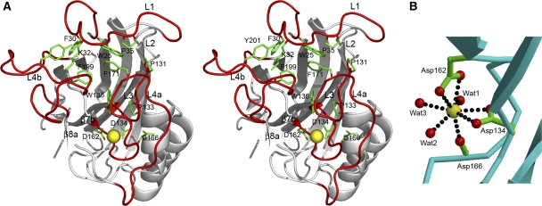
Figure 2.
Calcium-binding site of mindin-FS. (A) Stereo view of the top of mindin-FS. Loops L1, L3 and L4 are red. Conserved residues within these loops that participate in key interloop interactions, or in coordinating the Ca2+ ion (yellow), are green. (B) Coordination geometry of the Ca2+ ion. The calcium is ligated by Asp134, Asp162, Asp166, and three water molecules (Wat1–Wat3). Oxygen and carbon atoms are red and green, respectively.

