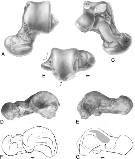
Fig. 3.
NMMP-39, left talus from Segyauk kyitchaung, in dorsal (A), posterior (B), plantar (C), lateral (D), and medial (E) views; the schematic drawings at the bottom (F and G) are derived from the lateral (D) and medial (E) views, respectively. The arrow shown in the posterior view (B) indicates the midtrochlear position of the flexor fibularis groove. The arrow shown in the schematic drawing of the medial view (G) indicates the elevated position of dorsally limited talo-tibial facet (gray claw-shaped area). Drawings (A-C and F-G) and plate conception are from L. Meslin (Institut des Sciences de l'Évolution de Montpellier). (Scale bars, 1 mm.)

