
Figure 2.
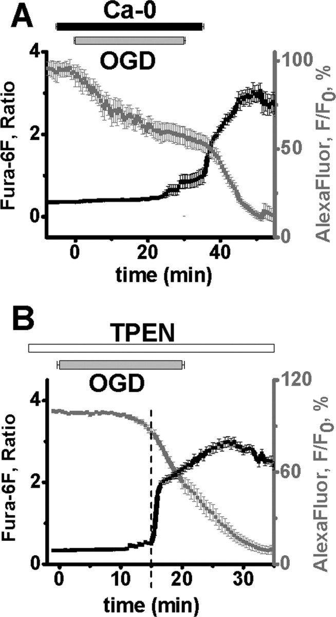
Zn2+ and Ca2+ contribute to OGD-induced damage of CA1 pyramidal neurons. A, Ca2+ removal during OGD prevents the Ca2+ deregulation. CA1 neurons were loaded with fura-6F (black) and AlexaFluor-488 (gray). Slices were superfused with Ca2+-free ACSF for 5 min before, during, and for 5 min after a 30 min episode of OGD. In cells exposed to OGD for 30 min, Ca2+ deregulation occurred almost immediately (1.5 ± 0.6 min, n = 8) after restoration of Ca2+ to the media, and as previously observed with Ca2+ containing ACSF, was accompanied by an abrupt acceleration in the rate of AlexaFluor-488 fluorescence loss, indicative of a terminal loss of membrane integrity. The slow loss of AlexaFluor-488 fluorescence throughout the OGD appeared to be largely attributable to enhanced cell swelling during zero-Ca2+ OGD. Individual responses (±SEM) have been aligned to the onset of Ca2+ deregulation, and the OGD bar shows the time of OGD exposure (±SEM), as above. B, TPEN significantly delays OGD-evoked Ca2+ deregulation. Neurons were loaded with fura-6F (black) and AlexaFluor-488 (gray), and exposed to TPEN (40 μm) before and during OGD exposure in normal Ca2+ containing ACSF (n = 12). Fura-6F signal increases and loss of membrane integrity (indicated by loss of AlexaFluor-488 fluorescence) (±SEM) still occurred, but were significantly delayed compared with responses in normal ACSF (see Fig. 1C). Individual responses have been aligned to the onset of Ca2+ deregulation, and the OGD bar shows the time of OGD exposure (±SEM), as above.